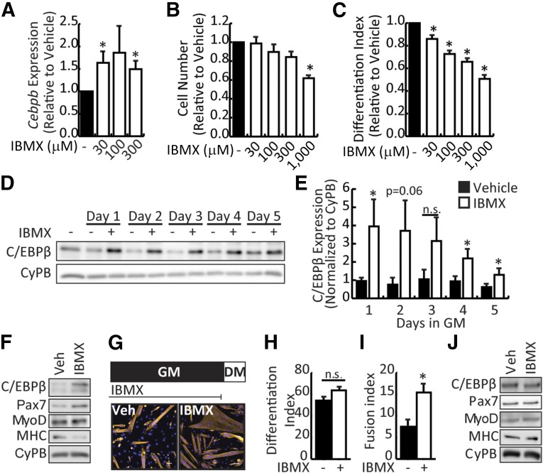
Figure 1.
IBMX stimulates expression of C/EBPβ in myoblasts. (A): Cebpb expression in primary myoblast cultured in 0, 30, or 300 μM IBMX for 24 hours in GM. (B): Cell numbers after culture of primary myoblasts in IBMX under growth conditions for 2 days. (C): Differentiation index (number of nuclei in MyHC+ cells/total nuclei) of primary myoblasts cultured for 2 days in differentiation medium in the absence or presence of IBMX. (D): Representative Western blot of C/EBPβ expression in primary myoblasts treated with 30 μM IBMX for up to 5 days in GM. CyPB is a loading control. (E): Quantification of C/EBPβ expression normalized to cyclophilin B. (F): Western analysis of myogenic marker expression in primary myoblasts cultured in vehicle or IBMX for 5 days under growth conditions and then induced to DM for 2 days in the continual absence or presence of IBMX. (G): Primary myoblasts were cultured in vehicle or IBMX for 5 days in GM, harvested, replated, and induced to DM in the absence of IBMX (top). Cells were fixed and stained for myosin heavy chain (yellow) and 4′,6-diamidino-2-phenylindole (blue). Representative pictures are shown (bottom). Scale bar = 50 μm. MyHC+ nuclei were counted to assess (H) differentiation index (as in C) and (I) fusion index (number of nuclei in MyHC+ cells/number of myotubes) for cells treated and differentiated as in (G). (J): Western analysis of myogenic marker expression from myoblasts cultured as in (G). All data are presented as mean ± SEM (n = 3; ∗, p < .05). Abbreviations: C/EBPβ, CCAAT/enhancer-binding protein β, CyPB, cyclophilin B; DM, differentiate; GM, growth medium; IBMX, isobutylmethylxanthine; n.s., not significant; Veh, vehicle.

