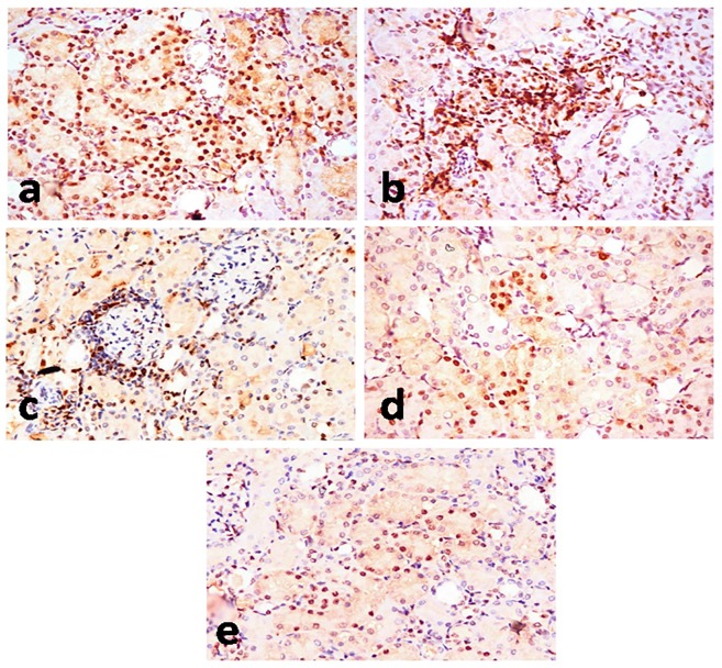
Fig 6. PCNA-immunohistochemical staining of kidney section of rats.
Kidney sections of PDC-treated group (a, b, c) showing significant increase of PCNA-positive renal tubular cells (a), PCNA-positive proliferating mononuclear cells in interstitial tissue (b), and PCNA-positive proliferating mononuclear cells in the periglomerular area (c). Kidney sections of PDC-Lf (200 mg/kg) treated rats (d) showing decrease of PCNA-positive renal tubular cells, and PDC-Lf (300 mg/kg) treated rats (e) decrease of PCNA-positive renal tubular cells. (Immunohistochemical staining of PCNA, X400).

