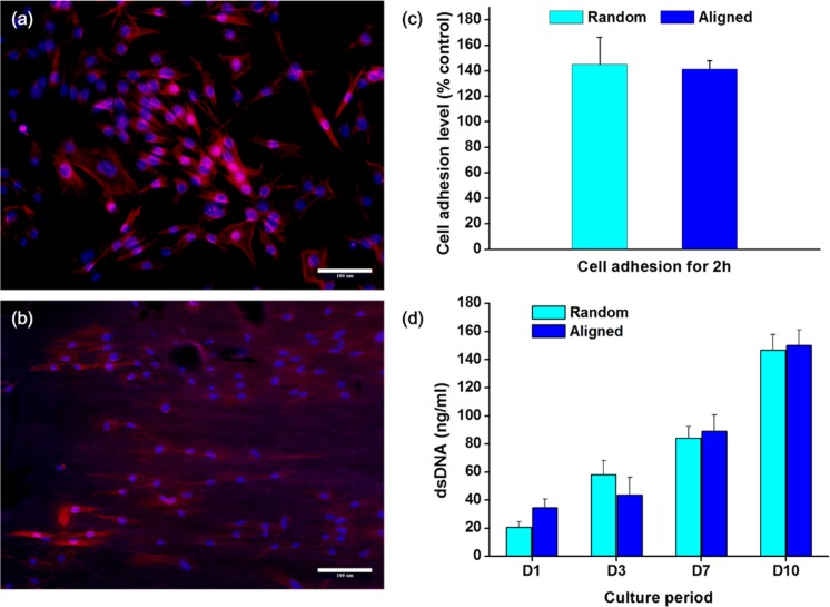
Fig 3. Effects of the nanofiber membrane on the initial cellular adhesion behavior.
Fluorescent images of cell attachment on (a) random and (b) aligned nanofiber. PDL cells recognize the underlying nanofiber alignment, conforming the shape of cell spreading to the nanofiber orientation (Magnification x200, scale bar 100 μm). (c) Cell attachment level, and (d) subsequent proliferation (*p < 0.05, by student t-test).

