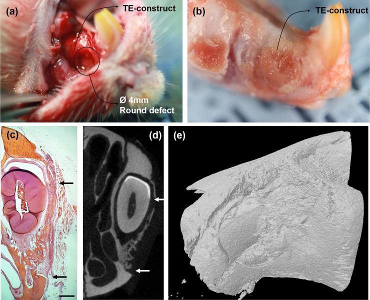
Fig 9. Illustrative images showing the PDL defect models used in this study.
(a) Photograph of rat premaxillary operation field. Note the dimensions of the defect used to produce standardized 4 mm diameters round full-thickness defects on the lateral surface of premaxilla bone. Two defects were created on one animal and were covered with tissue-engineered construct. (b) Harvested specimens of rat premaxillary operation field after sacrifice. (c) Representative histology image of HE staining of new bone tissue formed within the defect at 4 weeks (black arrow: defect margins) (Magnification x40, scale bar 500 μm). (d) 2D and (e) 3D μCT images. The original outline of the 4 mm defect is clear (white arrow).

