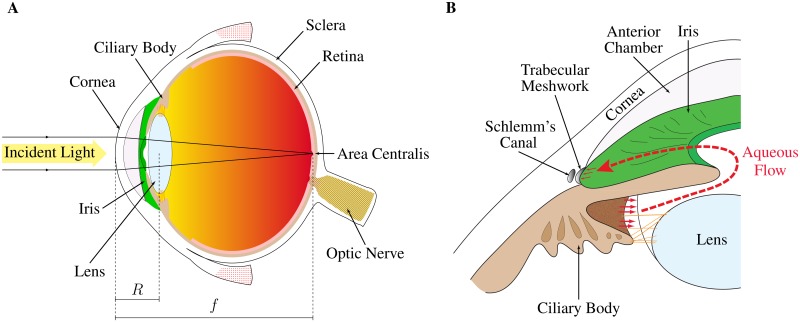
Fig 1. Schematic diagrams of a human eye (A) and the conventional aqueous flow pathway (B).
The human eye (A), which is fairly representative of the vertebrate eye, is composed of concentric layers of tissue enclosing a fluid filled chamber. Light is scattered towards the back of the eye by the cornea and lens. Phototransduction is carried out in the retina. Most of the light is focused on an area centralis, which here coincides with the fovea. The intraocular pressure is maintained by the equilibrium between the formation of aqueous humour and the resistance to its outflow from the eye. Produced by the ciliary body, the aqueous humour flows around the iris into the anterior chamber and is drained through the trabecular meshwork and Schlemm’s canal (B).

