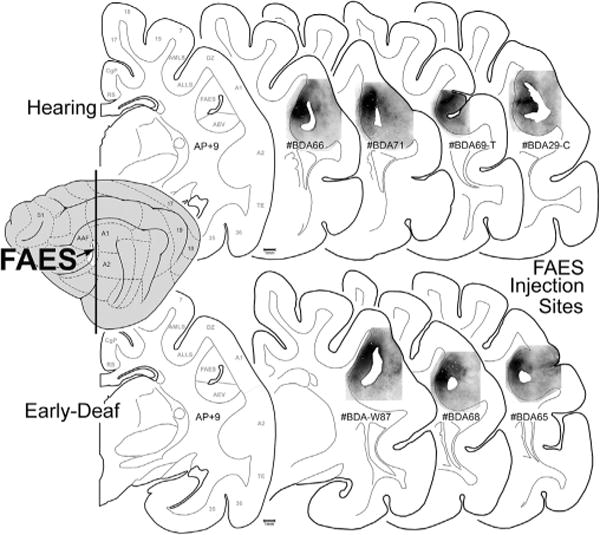
Fig. 2.

FAES tracer injection site summary. The lateral view of the cat cortex (left) indicates the location of the FAES (colored white at arrow) in relation to other cortical fields. The vertical line represents the approximate anterior-posterior (A–P) level of each of the coronal sections (to the right). The coronal section labeled AP + 9 is derived from Reinoso-Suárez (1961), with the functional subdivisions delimited by the gray lines (for abbreviation definitions see abbreviation table). Individual coronal sections illustrate the location of BDA tracer injection (blackened area) for the hearing (top row) and for the early-deaf cats (bottom-row) within the upper, medial bank of the sulcus corresponding to the position of the FAES region. Hearing case labeled #BDA69-T was comprised of an entire rostral-caudal series through the thalamus; hearing case labeled #BDA29-C was constituted by an entire rostral-caudal series through the cortex. All other cases had the full series of sections for both cortex and thalamus.
