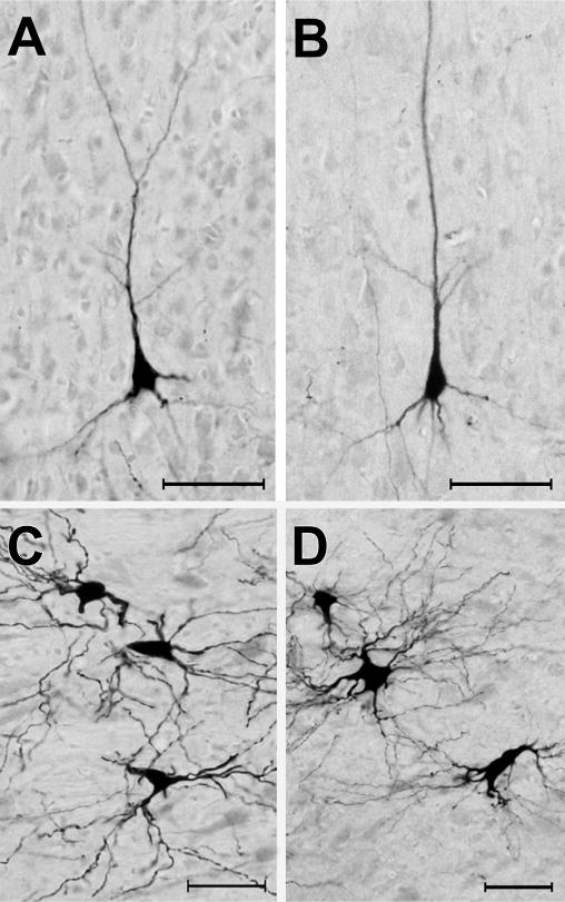
Fig. 3.

Photomicrographic examples of neurons retrogradely labeled by tracer (BDA) injection into the FAES. Panels A–B show BDA-labeled pyramidal neurons from area DZ in hearing (A) and early-deaf (B) animals (pial surface is toward the top). Panels C–D illustrated BDA-labeled neurons from the medial division of the MGB from hearing (C) and early-deaf animals. All scale bars = 50 μm.
