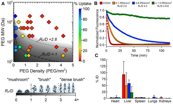
Figure 2.
The effect of surface PEG surface density on the uptake of polystyrene NPs by macrophages in vitro and circulation time in the blood in mice in vivo. (A) Phase diagram mapping NP uptake by differentiated human THP-1 cells as a function of PEG MW and coating density. A “dense brush” was most effective at reducing macrophage uptake in vitro. (B) The amount of NPs and NPs coated with PEG at various grafting densities circulating in the blood of mice over time, as observed using intravital microscopy. The RF/D values required for extended circulation time in vivo were increased compared to the PEG density required for reduced cell uptake in vitro. (C) Biodistribution of the various NP formulations 2 h after intravenous injection. Increased PEG surface density led to decreased accumulation in the liver. Adapted with permission from [51].

