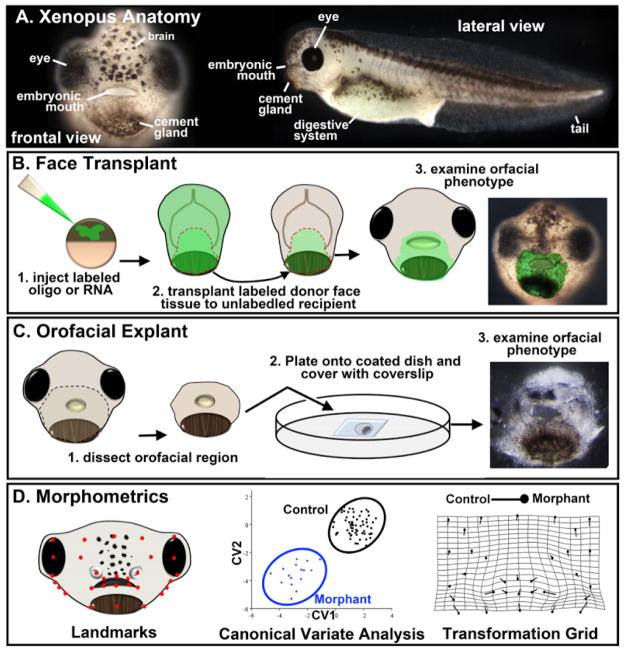
Figure 1.
Xenopus techniques for orofacial development. A) Frontal and lateral views of Xenopus at stage 40. B) Schematic showing the steps in performing a face transplant. Orofacial tissue is removed from a donor injected embryo and transplanted to the same region of sibling un-injected embryo. C) Schematic showing the steps in performing a face explant. Tissue is excised from the head and plated on a fibronectin coated glass bottom dish. D) Schematic showing a representative example of morphometric analysis of the orofacial region in Xenopus. Landmarks are assigned coordinates which are then used to perform a canonical variate analysis. This analysis can statistically separate landmark locations from a normal and morphant (or chemically treated) embryo which can then be presented graphically or on a transformation grid.

