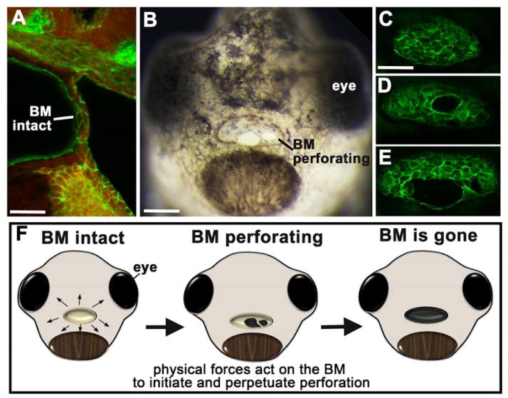
Figure 2.
Buccopharyngeal membrane rupture. A) Sagital section through the middle of the head showing the buccopharyngeal membrane. Cells are labeled with phalloidin (green) and the red is autofluorescence. Phalloin labels F-actin which is located along membranes of cells. scale bar=33 μm. B) Frontal view of a face as the buccopharyngeal membrane perforates or ruptures. scale bar=130 μm. C–E) Frontal views of phalloidin labeled buccopharyngeal membranes before (C) and during (D,E) perforation. scale bar = 80 μm. F) Schematic of embryonic faces at stage 40, showing the stages of buccopharyngeal membrane rupture and presenting the hypothesis that biomechanical forces are important for this process. Abbreviations: BM; buccopharyngeal membrane.

