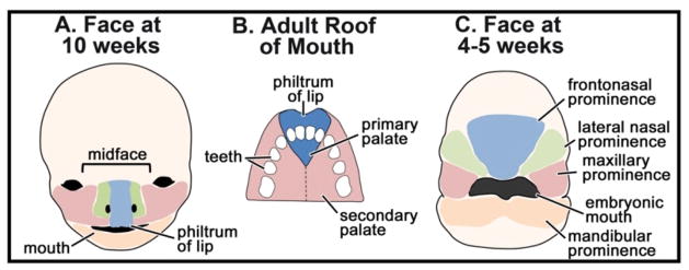
Figure 3.
Overview of orofacial development and primary palate anatomy. A) Schematic of the human fetal face at 10 weeks of development showing major anatomical features. The colored domains correspond to the facial prominences in (C) during embryonic development. B) Schematic of the adult palate showing the location of the primary verses secondary palate. C) Schematic of the face of a 4–5 week human fetus showing the location of the facial prominences.

