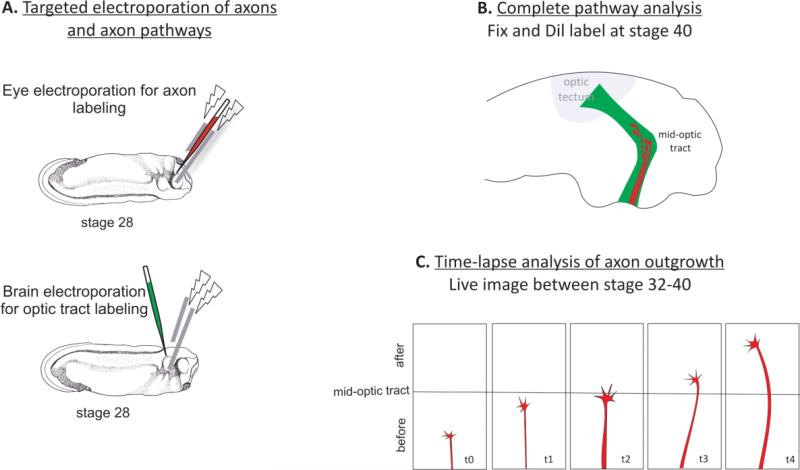
Figure 4. Xenopus visual pathway as a model of axon pathfinding.
Manipulation of genes in retinal axons and axonal pathways in the same embryo is possible via eye and brain targeted electroporation respectively. (A) Retinal axons and the neuroepithelium of the optic tract can be electroporated between stage 27 after axon initiation begins and stage 32 when the first retinal axons pass the optic chiasm and enter the optic tract in the brain. (B) For complete pathway analysis, embryos can be fixed at stage 40 and labeled with DiI or HRP. Labeled retinal pathway can be exposed via open brain preparation and pathfinding behaviors of axons can be analyzed. (C) Open brain preparations prepared in living embryos after stage 32 allows live imaging of retinal axons as they navigate through the optic tract in the brain. Time-lapse movies of axons can be recorded for 24h with 3min intervals.

