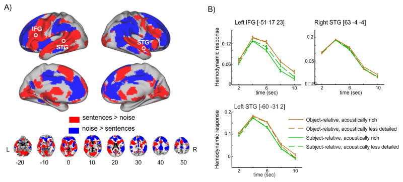
Figure 2.
A. Brain regions discriminating between intelligible sentence materials and unintelligible noise (1 channel vocoded speech). Significant voxels were identified using a whole-brain FIR model, subsequently color-coded according to the sign of the direct comparison of summed positive responses between conditions. B. fMRI intensities extracted from peak voxels located in left IFG, left STG, and right STG (circled in the rendering view). The x-axis depicts time relative to sentence onset. Error bars represent one standard error of the mean with between-subjects variance removed, suitable for within-subjects comparisons (Loftus and Masson, 1994).

