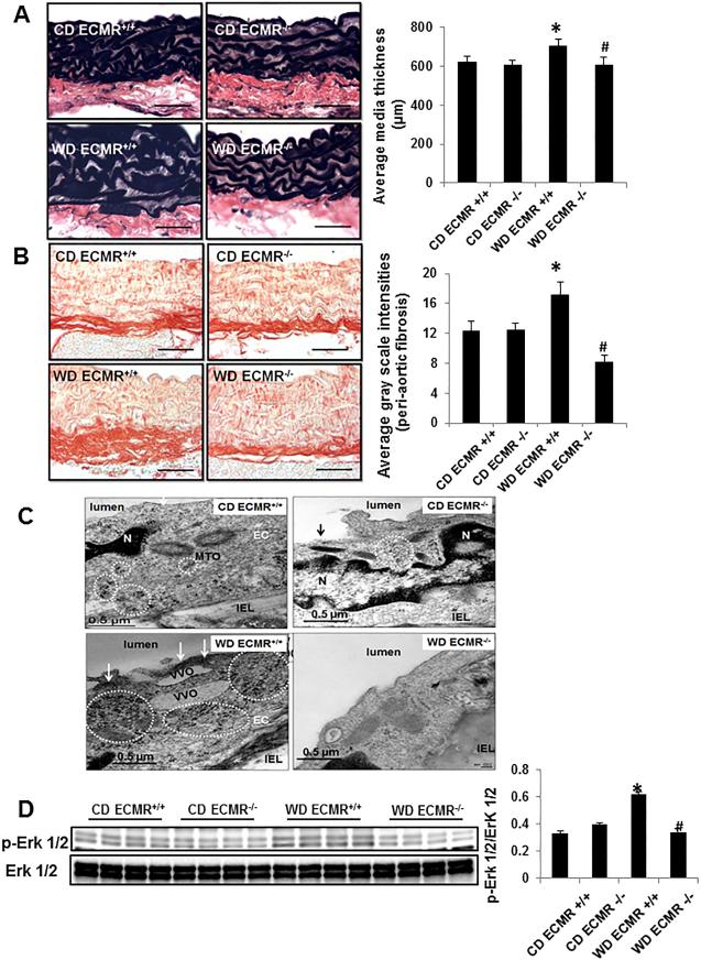
Figure 6. WD-induced aortic remodeling and ultrastructural abnormalities are prevented in in ECMR−/− female mice.
Representative micrographs show medial wall thickening staining with Verhoeff-Van Gieson (A) and periaortic fibrosis staining with picrosirius red staining (B). (C) ECMR−/− prevents WD-induced increases in thickened electron dense plasmalemma and free ribosomes, which contributed to EC stiffness. MTO= microtuble organizing center; IEL = internal elastic lamina; VVO=vesiculovacuolar organelles. Magnification X 10,000; bar = 0.5 μm. (D) ECMR−/− prevents WD-induced upregulation of p-Erk 1/2. n=4-6 per group. *P<0.05 compared with CD ECMR+/+; # P<0.05 compared with WD ECMR+/+.

