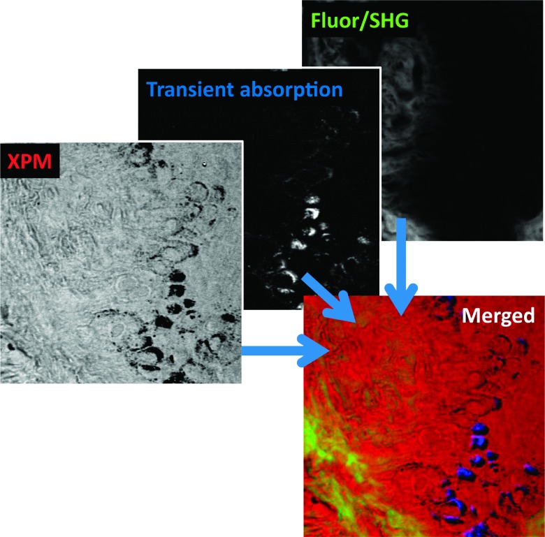
FIG. 12.
Images of the dermo-epidermal junction in a melanoma biopsy.68 Shown are XPMSS, transient absorption (tuned to visualize melanin), and combined multiphoton autofluorescence and SHG. The merged image shows the comprehensive contrast through multimodal multiphoton imaging. Adapted with permission from Wilson et al., Biomed. Opt. Express 3, 854 (2012). Copyright 2012 Optical Society of America.

