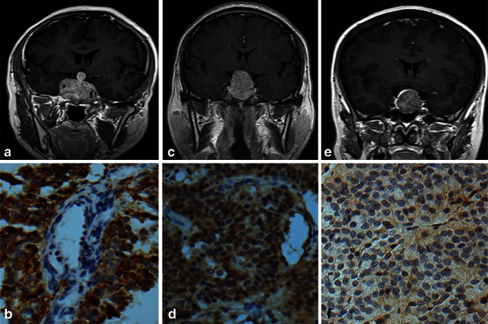
Fig. 1.
Coronal MR images with enhancement and endocan expression in the Knosp Grade 4 (a, b), 2 (c, d), and 0 (e, f) pituitary adenomas. In Grade 4 pituitary adenoma, ICA were entirely encased by tumor (a) and endocan was expressed in the cytoplasm of tumor cells and a few endothelial cells (b; IRS-TC = 12, IRS-EC = 1). Endocan expression was weaker in Grade 2 tumor which crossed the ICA center line but not the tangent line temporal side of blood vessel (c). Although endocan was expressed in tumor cells and endothelial cells, some cells were scattered in a negative expression (d; IRS-TC = 8, IRS-EC = 6). The Knosp Grade 0 adenoma did not cross the tangent line nasal side of blood vessel (e) and exhibited lower endocan reactivity (f; IRS-TC = 1, IRS-EC = 1). Magnification, ×400

