
Fig. 3.
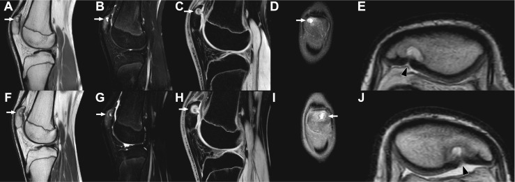
MRI of the right knee (a–e) and left knee (f–j) with sagittal proton-density (a, f), sagittal T2-weighted spectral attenuated inversion recovery (b, g), sagittal 3D water selective fluid (c, h), coronal proton-density (d, i), and magnified axial proton-density (e, j) slices shown. The lesion in the superolateral quadrant of both patellae, consistent with dorsal patellar defect, is demonstrated (arrows). Note the associated cartilage involvement with a slit-like defect/apparent discontuinity on both retropatellar surfaces (e, j, arrowheads). Also note the widespread surrounding high T2 signal in both patellae, consistent with bone marrow edema (b, g). MRI did not show any other abnormalities in either knee