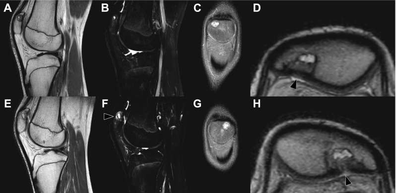
Fig. 5.

Eight-month follow-up MRI of the right knee (a–d) and left knee (e–h) with sagittal proton-density (a, e), sagittal T2-weighted spectral attenuated inversion recovery (b, f), oronal proton-density slices (c, g), and magnified axial proton-density views of the patellae (d, h) shown. The dorsal patellar defects are still present, but the surrounding bone marrow edema has decreased considerably (b, f) compared to 8 months earlier (Fig. 3). Only in the left knee some noteworthy bone marrow edema is still seen (f, arrowhead). Also note progressive “filling” of the dorsal defects with apparent (onset of) “closure” of the slit-like defects/discontuinities on both retropatellar surfaces (d, h, arrowheads) compared to 8 months earlier (Fig. 3). The patient was almost symptom-free at the time of this MRI examination
