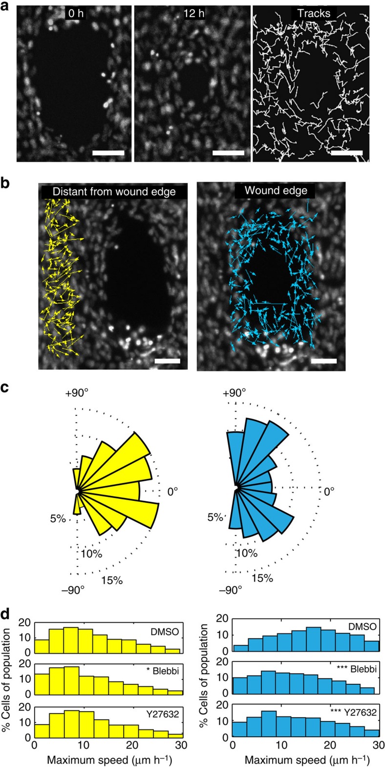
Figure 3. Gap closure is mediated by cell migration.
(a) Two frames of Hoechst labelled 3T3 fibroblasts in a wounded microtissue taken at 0 and 12 h of a time-lapse sequence during closure. Intermittent tracks of nuclei show non-correlated migration between neighbouring cells. (b) Vector fields showing net displacements for cells located at the wound edge versus 100 μm distant of the wound edge during closure. (c) Windrose plots displaying the frequency of migrating cells (%) versus the radial migration angle (0° means displacement towards the gap, 90° means tangential movement). (d) Histograms showing the maximum speed of cells distant of the wound edge when treated with dimethyl sulfoxide (DMSO), Blebbistatin and Y27632 (N=700–1,000 cells from 4 microtissues per condition, Kruskal–Wallis test, post-hoc test: Dunn Method For Joint Ranking with DMSO condition as control, *P<0.05 and ***P<0.001). Scale bars, 100 μm.

