
Figure 3.
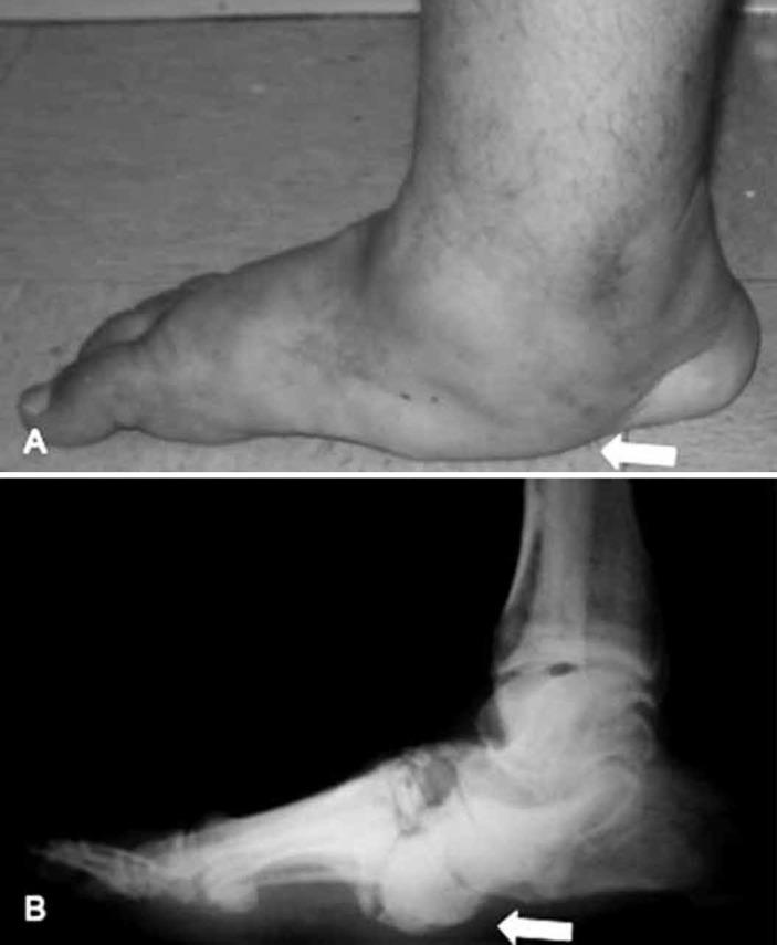
Profile of the right foot of the same patient as in Figures 1 and 2 after completing the treatment with TCC, showing the plantigrade position of the foot, now without detectable edema but with marked collapse of the medial arch (6A). Note also the prominence in the plantar region, characterizing the blotter appearance of the foot (arrow). Lateral radiograph of the foot (6B) with signs of bone consolidation, characteristic of Eichenholtz evolutional stage III. Note the persistence of a large bone prominence present in the plantar region of the midfoot (arrow), which is an area at risk of ulceration.
Source: SAME ISCMSP