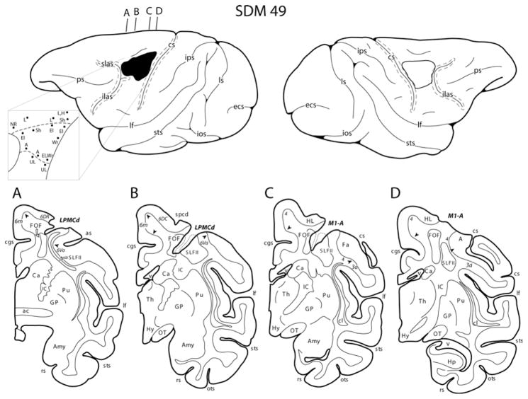
Fig. 2.
Line drawings of the lateral surface of the cerebral hemispheres of case SDM49 which received an M1 + LPMC lesion. Representative coronal sections through the lesion site are located immediately below the lateral views. The left hemisphere illustrates the location of the cortical lesion (blackened area) and the right hemisphere the location of the superimposed lesion (outlined area) that was used to calculate the respective gray and white matter lesion volumes using Nissl-stained tissue sections. Coronal panelsa–d are all through the lesion site on the left hemisphere. In each coronal section, the major region of extirpated cortex is identified by the bold italicized conventions. Pertinent Brodmann’s cytoarchitectonic areas are indicated on the coronal sections immediately below or within the gray matter (small italicized numbers/letters), and the respective boundaries are identified by the arrow heads. Identified in the subcortical white matter region are the putative locations of the fronto-occipital fasciculus (FOF) and superior longitudinal fasciculus subcomponent II (SLFII). The pullout illustrates the microstimulation map on the left hemisphere. On the map, each black dot represents a stimulation point with the affiliated movement(s) observed following stimulation. A arm of M1, ac anterior commissure, Amy amygdala, as spur of arcuate sulcus, Ca caudate nucleus, cgs cingulate sulcus, cl claustrum, cs central sulcus, ecs ectocalcarine sulcus, El elbow, GP globus pallidus, Fa face of M1, H hip, Hp hippocampus, HL hindlimb of M1, Hy hypothalamus, ic internal capsule, ilas inferior limb of arcuate sulcus, ios inferior occipital sulcus, ips intraparietal sulcus, L leg, lf lateral fissure, LPMCd dorsal lateral premotor cortex, ls lunate sulcus, M1 primary motor cortex, NR no response, OT optic tract, ots occipito-temporal sulcus, ps principal sulcus, Pu putamen nucleus, rs rhinal sulcus, Sh shoulder, slas superior limb of arcuate sulcus, spcd superior precentral dimple, sts superior temporal sulcus, Th thalamus, UL upper lip, v ventricle, Wr wrist

