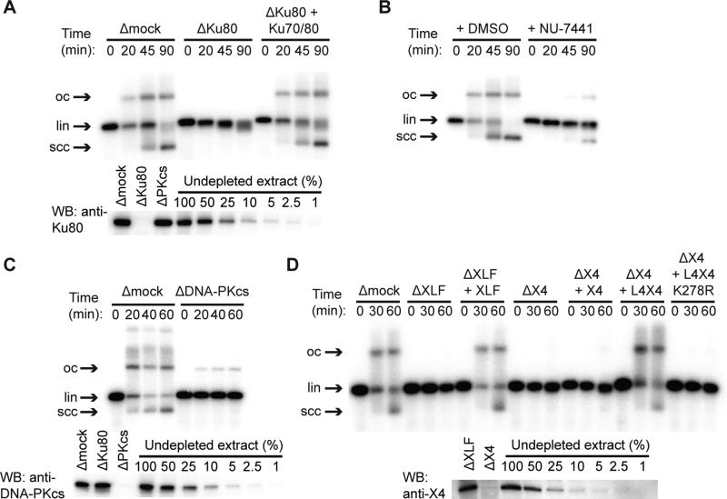
Figure 1. End Joining in Xenopus Egg Extract Depends on Classical NHEJ Factors.
(A) Inhibition of end joining by immunodepletion of Ku70/80 with αKu80 antibody, and rescue with recombinant X. laevis Ku70/80. lin, linear DNA substrate; scc, supercoiled closed-circular products; oc, open-circular products. In other conditions, Ku immunodepletion selectively inhibited circularization, as previously reported (Fig. S1C) (Labhart, 1999; Di Virgilio and Gautier, 2005).
(B) Inhibition of end joining by the DNA-PK inhibitor NU-7441.
(C) DNA-PKcs immunodepletion.
(D) Immunodepletion of XLF (ΔXLF) or XRCC4 (ΔX4) and rescue with recombinant X. laevis XLF, XRCC4 (X4), wild-type LIG4:XRCC4 (L4X4), or catalytically inactive LIG4K278R:XRCC4 (L4X4 K278R). Lower panels in (A), (B), and (D) are western blots of immunodepleted extract with indicated antibodies. Uncropped blots are shown in Fig. S1F-H. XLF was not clearly visible in western blots of extract, but immunoprecipitated XLF was detected by western blotting and mass spectrometry (Fig. S1J-K).
See also Figure S1.

