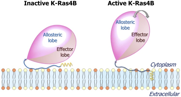
Figure 10.
Diagrams representing the inactive (left) and active (right) states of K-Ras4B at the anionic membrane. The balloon represents the K-Ras4B catalytic domain, the blue thread tied to the balloon represents the HVR, and the yellow sawtooth represents the farnesyl. In the inactive state, the membrane-interacting HVR hauls the effector lobe to the membrane surface, burying the effector-binding site. In the active state, the catalytic domain liberates the HVR, exposing the effector-binding site and fluctuating reinlessly.

