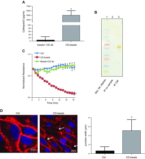
Figure 2.
The 30–100 kDa fraction contains CD, which alters the EC barrier. A) CD levels associated with agarose beads as measured by ELISA following IP. *P = 0.02, significantly greater than beads plus antibody (ab) without exposure to the 30–100 kDa fraction. B) Silver-stained gel. Mol. Wt. (molecular mass) marker is in lane 1, IP no sample is in lane 2, and IP CM is in lane 3. C) Representative ECIS tracings of HRECs treated with CD beads demonstrate significantly decreased resistance in comparison to cells treated, beads plus CD antibody (ab), or Ctrl. *P = 0.006, significantly less than cells grown in Ctrl. D) Representative image of VE-cadherin immunofluorescence of HRECs grown in Ctrl and in the presence of CD beads. White arrows indicate gaps between adjacent cells treated with CD beads. Quantitation demonstrates significantly increased gap width (micrometers) between adjacent cells. Data are means ± sd. *P = 0.0001, significantly greater than cells grown in Ctrl.

