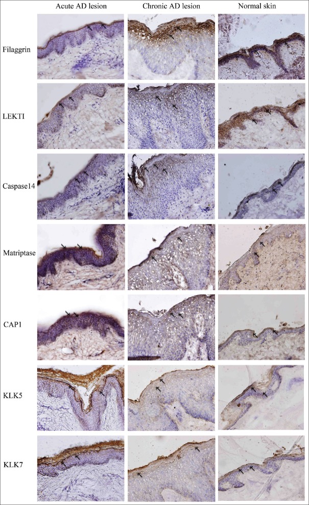
Figure 1.
Expression models of filaggrin and proteases involved in filaggrin processing in acute and chronic lesions of atopic dermatitis patients. Skin sections were stained for filaggrin, LEKTI, caspase14, matriptase, CAP1, KLK5, and KLK7. Filaggrin, LEKTI, and caspase14 expressions were significantly decreased, whereas matriptase, CAP1, KLK5, and KLK7 expressions were increased in AD lesions, as compared to normal controls. This phenomenon was more pronounced in the skin from patients with acute lesions (DAB staining, Original magnification ×100, the arrows point to the stained positive cells), (n = 3/three experiments in duplicate). AD: Atopic dermatitis; LEKTI: Lymphoepithelialkazal-type-related inhibitor; CAP1: Channel-activating serine protease 1; KLK5: Kallikrein 5; KLK7: Kallikrein 7.

