
Figure 1.
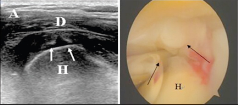
Articular-side partial-thickness tear sonographic image shows that anechoic fluid fills in the region of articular-sided tendon (white arrows, left image) while bursal-sided tendon is still relatively intact. This kind of injury is considered partial-thickness tear at the supraspinatus tendon articular-side. The result is confirmed with arthroscopic (black arrows, right image). H: Humeral head; D: Deltoid.