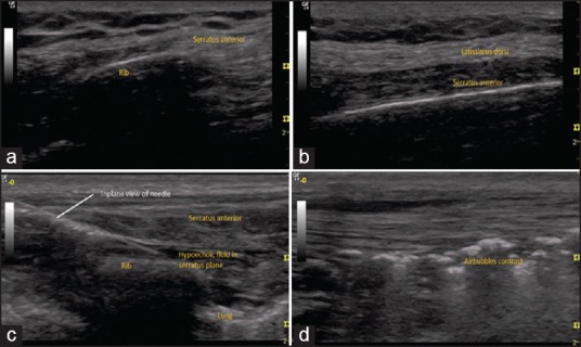
Figure 1.

(a) Ultrasound image of incomplete serratus muscle belly in anterior axillary line (b) Ultrasound image of complete belly formation in posterior axillary line (c) Ultrasound image of in-plane view of needle and hypoechoic fluid in-plane (d) Ultrasound image of hyperechoic air bubbles in-plane
