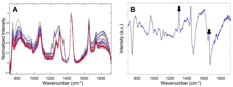
Figure 6.
Noninvasive Raman spectroscopy accurately detects WATi in a mouse model of obesity. (A) Transcutaneous Raman spectra from inflamed (red) and noninflamed (blue) murine mammary WAT (n = 5/group). (B) Diagnostic PC highlighting Raman peaks at 1264 and 1652 cm−1 (see arrowed features), which were found to be diagnostic in all other datasets.

