Figure 2.
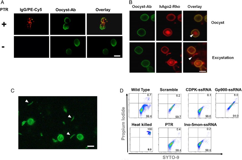
Cryptosporidium transfection and viability assays. A, Fluorescent immunoglobulin G (IgG) labeled with phycoerythrin (red) was encapsulated within protein transfection reagent (PTR) and then used to transfect Cryptosporidium sporozoites within oocysts. Oocysts were incubated with anti-Cryptosporidium oocyst antibody-FITC (Oocyst Ab-FITC; green). IgG was only introduced into samples treated with PTR (top). No signal was detected in the parasites with samples incubated only with IgG (bottom). B, PTR was loaded with labeled human Argonaute 2 (hAgo2)–NHS-rhodamine (red) and then used to transfect oocysts. Cyst wall was stained with oocyst antibody (green). hAgo2 was detected in oocysts (white triangle; top) or in released sporozoites after excystation (white triangle; bottom). C, We stained oocysts with the vital dye carboxyfluorescein succinimidyl ester (CFSE; green). Stained and motile sporozoites were observed (white triangles). White bar, 5 µm. D, Viability was quantified by flow cytometry for untransfected parasites (wild type), heat-killed parasites, parasites treated with empty PTR, or parasites transfected with complexes of hAgo2 preloaded with scramble single-stranded RNA (ssRNA), calcium-dependent kinase 1 (CDPK)–ssRNA, inosine-5-monophosphate dehydrogenase (Ino-5mon)–ssRNA, and glycoprotein (Gp900)–ssRNA.

