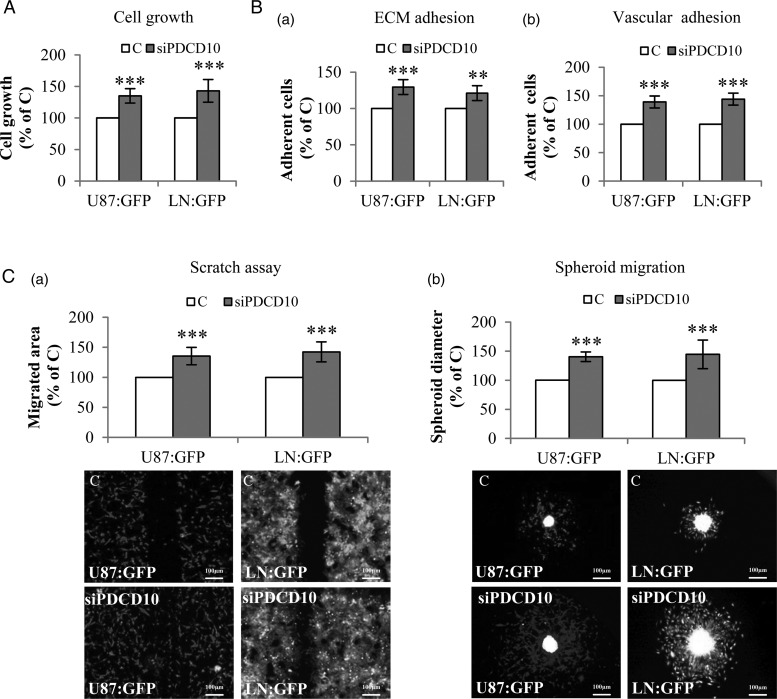
Fig. 1.
Direct co-culture of glioblastoma (GBM) cells with PDCD10-silenced human umbilical vein endothelial cell (HUVEC)-activated tumor phenotyping. HUVECs were transfected with either siPDCD10 or control siRNA (C) and used for direct co-culture with GBM cells (U87:GFP or LN:GFP) after 72 hours of the transfection. (A) Endothelial silence of PDCD10 promoted GBM cell growth. The growth of U87:GFP or LN:GFP was detected as green fluorescent intensity at 485 nm of excitation wavelength and 535 nm of emission wavelength. (B) Endothelial silence of PDCD10 facilitated extracellular matrix (ECM) adhesion (a) and vascular adhesion (b) of GBM cells. The adherent U87:GFP or LN:GFP cells were quantified by detecting green fluorescent intensity of cells. (C) Endothelial silence of PDCD10 stimulated GBM cell migration. For scratch assay (a), the migrated area was calculated by subtracting the open area at zero hour from that measured at 24 hours after scratching. For spheroid migration assay (b), the diameter of spheroids was measured at 36 hours after seeding spheroids containing HUVECs and GBM cells. ** P < .01 and *** P < .001, compared to C. Scale bar: 100 µm.

