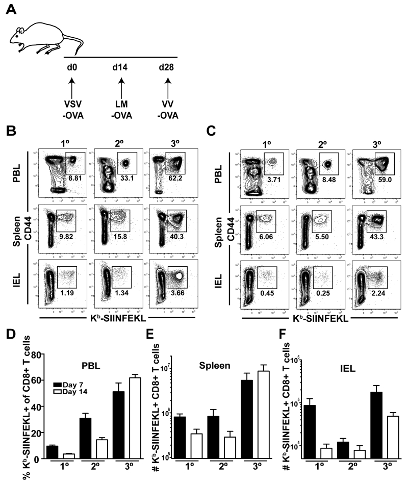
FIGURE 1. Short-boosting intervals generate large numbers of Ag-specific CD8 T cells.
(A) Short-boosting immunization regimen. Boosts occurred 14 days apart. (B, C) Peripheral blood lymphocytes (PBL), spleen, and small intestinal intraepithelial lymphocytes (IEL) were analyzed (B) 7 days or (C) 14 days after 1°, 2° or 3° boosting. Plots are gated on CD8 T cells. (D) Percent of CD44+ Kb-SIINFEKL+ CD8 T cells in PBL at day 7 (black) and 14 (white) after 1°, 2° or 3° boosting. (E-F) Number of CD44+ Kb-SIINFEKL+ CD8 T cells in spleen (E) and IEL (F) at day 7 (black) or day 14 (white) following 1°, 2° or 3° boosting. Data are representative of 2 experiments, N=3 mice per experiment.

