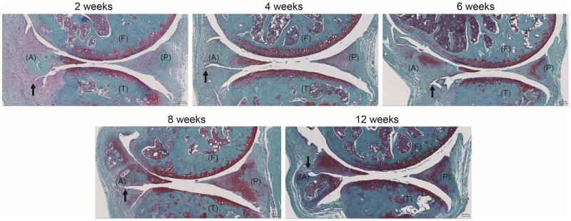
Figure 2.
Representative images of the surgical/injury DMM model of C57BL/6J mice knee joints (2-, 4-, 6-, 8- and 12-weeks post-destabilization) showing the femur (F), tibia (T) as well as anterior (A) and posterior (P) location of the menisci (Safranin O staining). The transection of the meniscotibial ligament (black arrow) intersects the medial menisci, leading to joint stability, meniscal degeneration, and development of OA in articular cartilage

