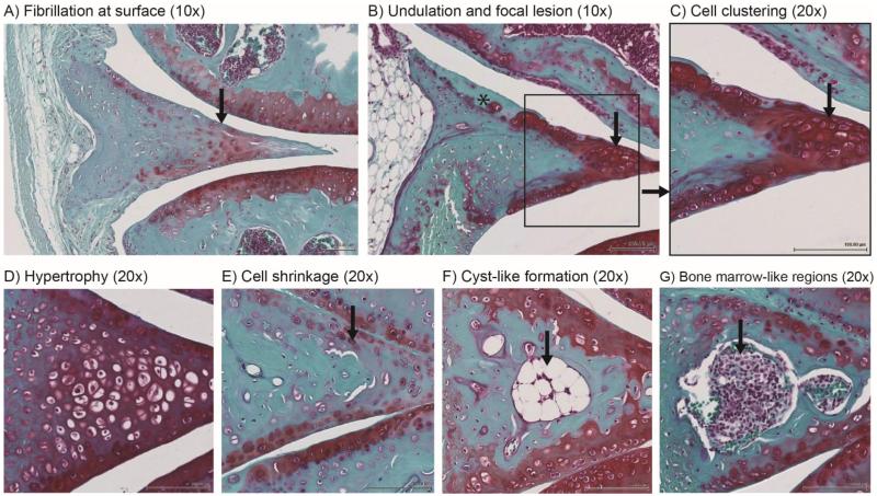
Figure 6.
Age-related abnormal changes to tissue structure, cells and extracellular matrix in aged mice (24 to 36 months old). Structural changes include (A) fibrillation (indicated by arrow) and (B) undulation mainly at the femoral aspect surface (asterisk indicates surface defects with faint or no Safranin O staining). Hypercellular changes included (C) cell clustering (shown in inset and indicated by arrow) and (D) hypertrophy. Hypocellularity was observed through (E) reduced cell density and cell shrinkage (indicated by arrow). (F) Cyst-like formation (indicated by arrow) and (G) bone marrow-like regions were also observed more frequently in aged mice (indicated by arrow).

