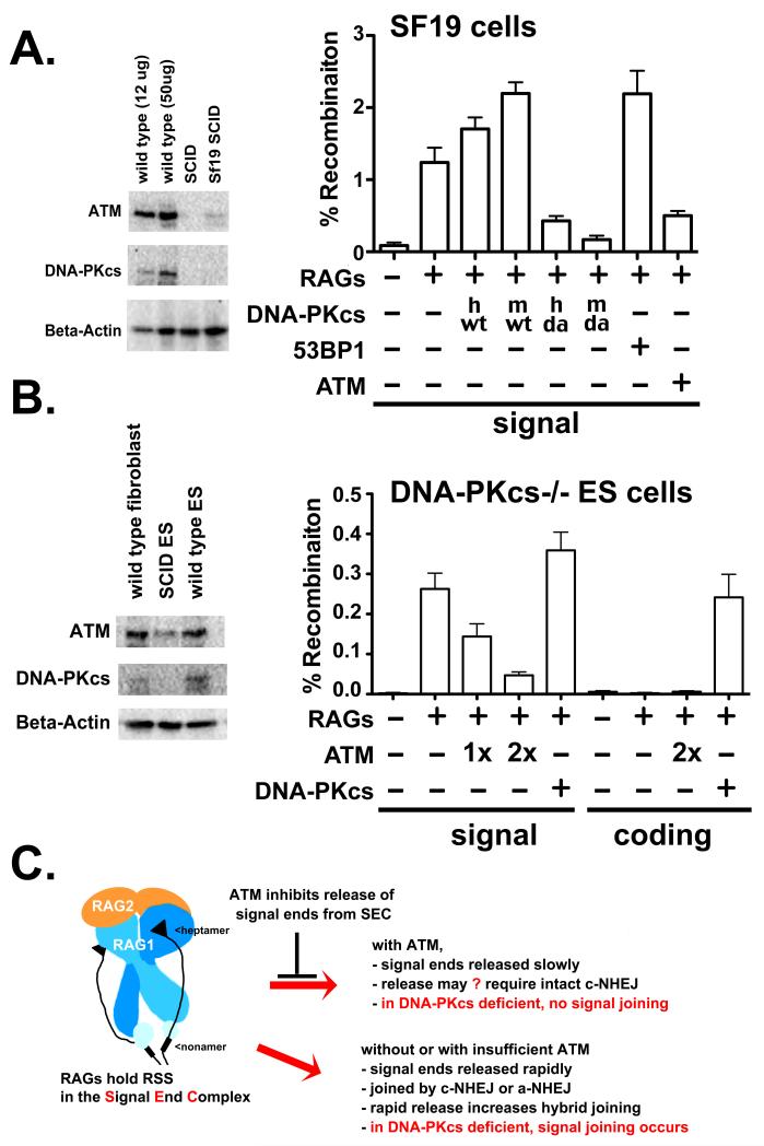
Figure 6. ATM over-expression suppresses signal joining in SCID mouse fibroblasts and DNA-PKcs deficient murine ES cells.
A. Left panel. Immunoblot analyses for ATM, DNA-PKcs or Beta-Actin expression of 12-50 μg of whole cell extracts from wild type or SCID ear fibroblasts, or the murine SCID fibroblast cell strain, Sf19. Right panel. Recombination percentage of the signal joint substrate 289/RFP/CFP in Sf19 cells transiently expressing RAG proteins and either no DNA-PKcs, human or mouse, wild type or mutant DNA-PKcs (vector, hwt, mwt, hD3922>A, mD3922>A,), 53BP1 or ATM. Cells were analyzed by flow cytometry as in Fig. 1. B. Left panel. Immunoblot analyses for ATM, DNA-PKcs or Beta-Actin expression of 50 μg whole cell extracts from wild type mouse fibroblasts, and wild type or DNA-PKcs deficient murine ES cells strains. Right panel. Recombination percentage of the signal joint substrate pJH201 or coding joint substrate pJH290 in DNA-PKcs deficient murine ES cells transiently expressing RAG proteins and varying levels of ATM, vector control, or DNA-PKcs as indicated. C. Proposed model. SEC structure is based on studies of Ru et al. (77).

