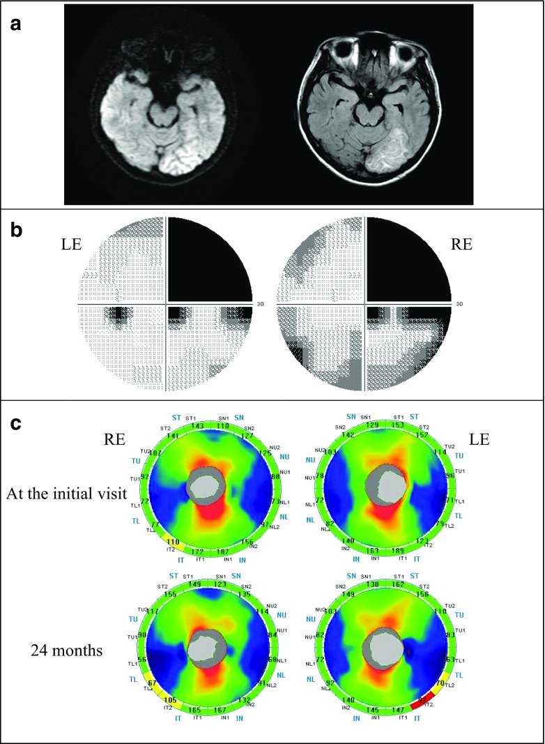
Fig. 4.
Patient with right homonymous hemianopia (case 4). a A 39-year-old woman with an infarction in the left posterior cerebral artery territory. Left: a diffusion weighted image (DWI), Right a fluid attenuated inversion recovery (FLAIR) image. DWI and FLAIR images showed an ischemic stroke on the left occipital lobe which was represented as a high-intensity lesion 3.5 months after the onset. b Humphrey visual fields showed right homonymous hemianopia denser superiorly. RE right eye, LE left eye. c A Significance map of the cpRNFL thickness. Top: at the initial visit, Bottom: 24 months after the onset. RE right eye, LE left eye. At the initial visit, cpRNFL thinning was observed in the inferior temporal sector of the RE. There was no significant change in the LE. After 24 months, the thinning of RE was progressive and thinning sectors of LE corresponded to the visual field defect

