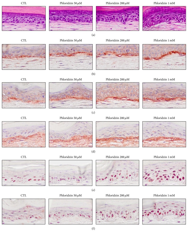
Figure 2.
Histologic findings for PZ-treated SEs. SEs were constructed and then incubated in the presence of PZ (0, 50, and 200 μM or I mM). Sections of SEs were stained with hematoxylin and eosin and analyzed by immunohistochemical staining ((a): H&E staining, (b): integrin α6, (c): integrin β1, (d): type IV collagen, (e): p63, and (f): PCNA).

