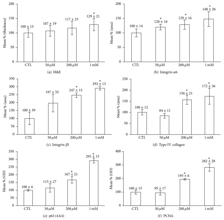
Figure 3.
Comparison of epidermal thickness, staining intensity, and the numbers of p63- and PCNA-positive cells. Immunohistochemical staining was analyzed quantitatively by using Image J software (National Institute of Health, Bethesda, Maryland, USA). The positivity of p63 and PCNA was measured as described in Section 2. ∗ P < 0.05 compared to contro.

