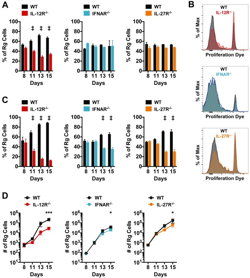
Figure 7. CD8+ T cell priming in the lung draining lymph node requires IL-12, while CD8+ T cell expansion in the lung depends on IL-12, type I IFN, and IL-27.
Equal numbers of retrogenic TB10.44-11-specific (TB10Rg) CD8+T cells were transferred into mice 7 days after low-dose aerosol infection with Mtb. (a) The percentage of total retrogenic (Rg) cells that were WT or KO for the indicated cytokine receptor in the mediastinal LN on days 8, 11, 13 and 15 following infection. (b) Histograms depicting the dilution of the proliferation dye efluor 450 in TB10Rg cells in the LN at day 11. Each group of samples (WT or KO) was concatenated into a single histogram. (c) The percentage of total retrogenic (Rg) cells that were WT or KO for the indicated cytokine receptor in the lungs at days 8, 11, 13 and 15 following infection. (d) Total number of WT and KO Rg cells detected in the lungs at the indicated time points. Each bar or point represents the mean ± SEM (n = 4-5 mice per group) *P < 0.05, **P < 0.01, ***P < 0.001, ‡P < 0.0001 (Holm-Šídák multiple comparisons testing following two-way ANOVA). Data are representative of two independent experiments.

