
Figure 2.
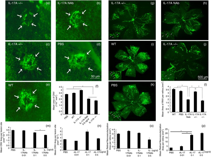
Immunofluorescent staining of retinas and choroidal flat‐mounts of models of mice with retinopathy of prematurity (ROP) and choroidal neovascularization (CNV). Interleukin‐17A‐deficient (IL‐17A−/−) and wild‐type (WT) C57BL/6 mice were subjected to hypoxic retinopathy and laser‐induced CNV as described in the Materials and methods section. Among them, the WT mice were divided into several groups at random, with one eye intravitreously injected with rIL‐17A or IL‐17A neutralizing antibody (NAb) and the other eye injected with PBS at postnatal day 12 (P12) for mice with ROP, and at days 1 and 7 after laser photocoagulation for mice with CNV. The retinas of mice with ROP were flat‐mounted and stained with FITC‐lectin at P17, while choroidal flat‐mounts from mice with CNV were performed at day 14 after photocoagulation as described (a–e: n = 10 mice/group; g–k: n = 10 mice/group). Dose–effect experiments of IL‐17 NAb and rIL‐17A were performed (m–p). The image‐pro plus software was used to quantify the area of NV (f and l). Statistics were analysed using the Student–Newman–Keuls method. Data were mean ± SEM; *P < 0·05.