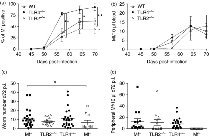
Figure 4.

Lack of TLR4 during Litomosoides sigmodontis infection increases the percentage of Mf+ mice. WT, TLR2−/− and TLR4−/− BALB/c mice were naturally infected with L. sigmodontis and from day 45, mice were surveyed for the presence of peripheral Mf. (a) Symbols represent the mean percentage ± SEM of mice that were Mf+ within the different strains. (b) Symbols show the mean percentage ± SEM of Mf number per 10 μl blood in individual mice. On day 72, (c) worm burden and (d) peripheral Mf numbers were determined in individual mice. (a) Data from four independent infection experiments (n = 45 WT, n = 25 TLR2−/− and n = 32 TLR4−/−). (b–d) Values show data from three independent infection studies (n = 20 WT Mf+, n = 11 WT Mf–, n = 12 TLR2−/− Mf+ mice and n = 22 TLR4−/− Mf+ mice) and correlate to the data described in Figs 5, 6, 7. Asterisks indicate significant differences (analysis of variance or Student's t‐test) between the groups indicated by the brackets (*P < 0·01).
