
Figure 8.
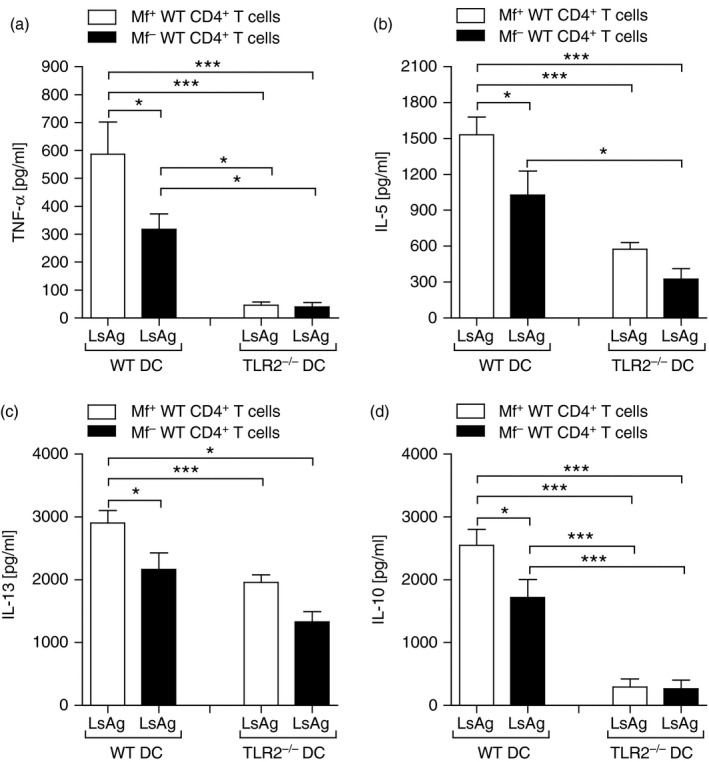
Lack of TLR2 on APC reduces filarial‐specific CD4+ T‐cell responses from WT infected mice. On day 72 post infection (p.i.), CD4+ T cells from Mf+; white bars or Mf– (black bars) infected BALB/c mice were isolated by flow cytometry and co‐cultured with DC from WT or TLR2−/− BALB/c mice. Cell cultures were then stimulated with 50 μg/ml Litomosoides sigmodontis antigen (LsAg) for 72 hr and thereafter, levels of TNF‐α (a), IL‐5 (b), IL‐13 (c) and IL‐10 (d) were determined in the culture supernatant by ELISA. From three independent infection studies, bars show mean ± SD of three co‐culture assays using isolated T cells pooled from three or four mice per group. Asterisks indicated significant differences (analysis of variance or Student's t‐test) between the groups indicated by the brackets (*P < 0·01, ***P < 0·001).