
Figure 2.
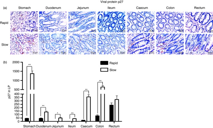
Detection of the viral protein p27 (brown) in the gastrointestinal (GI) tracts of rapid and slow progressors. (a) Representative images (400 ×) of the GI tract stained for p27 antigen (light red) are shown. (b) Quantitative analysis of p27 (* : P ≤ 0.05, ** : 0.05 < P ≤ 0.01).