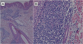
Figure 2.

(A) dense lymphocytic infiltrate surrounding dermal vessels with focal involvement of the wall without epidermotropism or basal layer changes. Adjacent ectatic lymphatic vessels were also present. Hematoxylin‐eosin stain; original magnification x 100 and (B) the inflammatory infiltrate is predominantly formed by T lymphocytes (CD3+/CD4+/CD8+), few histiocytes and plasma cells. It involves the full thickness of the dermis with Jessner‐type pattern around vascular plexuses, adnexal structures and nerve endings. Rare extravasated red blood cells were also present. Hematoxylin‐eosin stain; original magnification x 250
