
Figure 1.
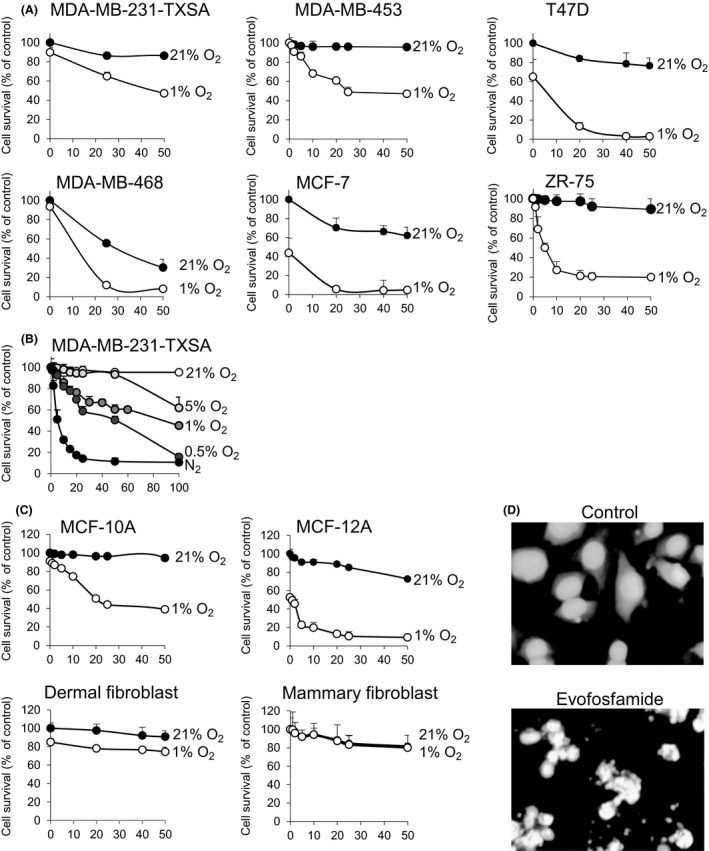
Activity of evofosfamide against breast cancer, epithelial cells, and fibroblasts in vitro. (A) Six breast cancer cell lines were seeded in 96‐well plates at 1 × 10⁴ cells per well and treated with increasing doses of evofosfamide in normoxic (21% O2) and hypoxic (1% O2) conditions for 48 h. (B) The breast cancer cell line MDA‐MB‐231‐TXSA was treated with an increasing dose of evofosfamide under normoxic (21% O2) and in various hypoxic conditions from 0% to 5% O2 for 24 h. (C) Evofosfamide reduced cell viability in both epithelial breast cell lines MCF‐10A and MCF‐12A selectively under hypoxic conditions (1% O2) 48 h after treatment. Dermal and mammary fibroblasts were relatively resistant to evofosfamide under the same conditions. Cell viability of all cell lines was assessed by crystal violet staining. (D) DAPI nuclear fluorescence stain of untreated MDA‐MB‐231‐TXSA cells showing nuclei homogenously stained. Cells treated with 50 μmol/L evofosfamide under hypoxic conditions for 24 h exhibit changes in the nuclei consistent with the induction of apoptosis. Data points show means of quadruplicate results from a representative experiment, repeated at least twice and presented as the mean ± SD of quadruplicate wells and expressed as a percentage of the number of control cells.