
Figure 3.
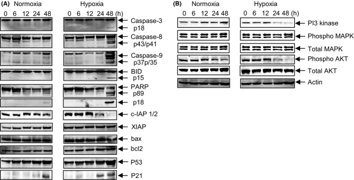
Apoptotic signaling of evofosfamide as a single agent against MDA‐MB‐231‐TXSA cells. (A) MDA‐MB‐231‐TXSA‐TGL cells were seeded at 2 × 10⁶ per T25 flask and were treated with evofosfamide at 50 μmol/L under normoxic (21% O2) and hypoxic (1% O2) conditions. Cells were then lysed and protein lysates were collected at 0, 6, 12, 24, and 48 h after treatment. Cell lysates were analyzed by polyacrylamide gel electrophoresis and transferred to PVDF membranes for immunodetection as described in Materials and Methods section. Evofosfamide treatment resulted in cleavage and prominent activation of the initiator caspase‐8 followed by caspase‐9, and caspase‐3 and the concomitant processing of BID and PARP protein, only at the later time point of 48 h, indicating that these changes were an effect rather than a cause of evofosfamide‐induced apoptosis. cIAP1/2 levels were significantly reduced with evofosfamide treatment, under hypoxic conditions at the 24‐ and 48‐h time points, whereas the levels of XIAP, BAX, and Bcl‐2 remained unchanged. (B) A robust reduction in PI3 kinase and phosphorylated AKT under hypoxic conditions after treatment with TH‐302 at 24 and 48 h. However, no significant changes in the levels of either phosphorylated or total MAPK were detected following treatment.