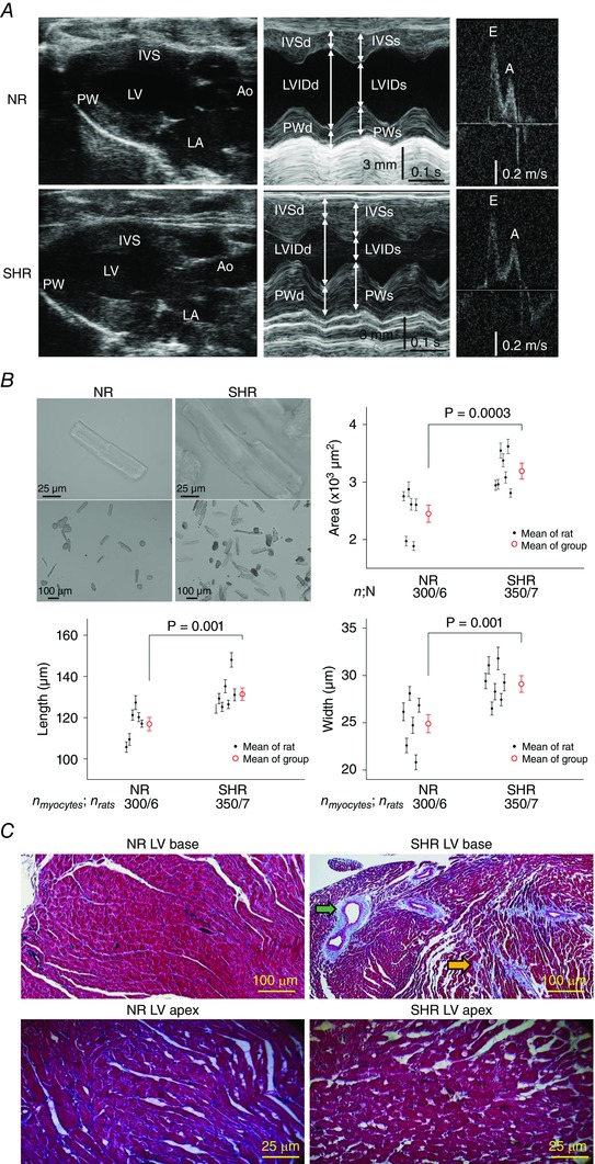
Figure 1. Cardiac remodelling in early hypertension of 5‐ to 6‐month‐old male SHR .

A, although both SHR and NR hearts had preserved LV systolic function, only SHR hearts manifested concentric LVH with global increased wall thicknesses seen on B‐mode (longitudinal, panel 1) and M‐mode (cross‐section, panel 2). Ao, aorta. E and A, peak velocities in early and late filling. B, SHR hypertrophy at the myocyte level is evident on bright‐field micrographs of representative myocytes and is confirmed by direct measurements of myocyte cross‐sectional area, length, and width. The mean ± SEM for each rat is represented by a small black circle with whiskers and, for each group by a large red circle with whiskers. n myocytes/n rats, number of myocytes/number of rats. C, although fibrosis is not apparent in the LV base from NR hearts (upper left) or the LV apex of both NR and SHR hearts (bottom), focal fibrosis (blue trichrome stain) is evident in the LV base from SHR hearts in the perivascular (green arrow) and interstitial (orange arrow) regions.
