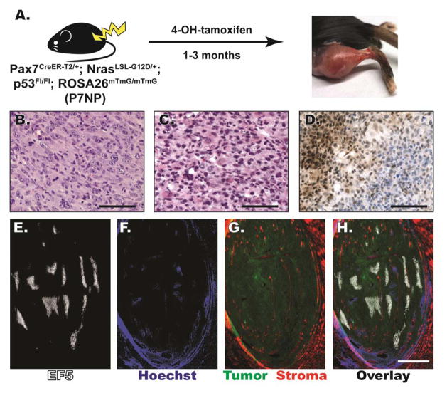
Figure 2. P7NP sarcomas were either UPS or RMS by histology, and had regions of HIF-1α accumulation and tumor hypoxia.
A) Schematic of novel Pax7Cre-ER-T2/+; NrasLSL-G12D/+; p53FL/FL; ROSA26mTmG/mTmG (P7NP) mouse model of soft tissue sarcoma generated by intramuscular 4-hydroxytamoxifen injection. (B, C) FFPE sections of primary soft tissue sarcomas from P7NP mice were stained with hematoxylin and eosin. B) P7NP sarcoma with histology resembling undifferentiated pleomorphic sarcoma (UPS). Scalebar = 100μm. C) P7NP sarcoma with histology resembling rhabdomysarcoma (RMS). Scalebar = 100μm. D) Representative FFPE sarcoma section stained with antibody against HIF-1α and signal-amplified with DAB. HIF-1α staining of sarcomas from P7NP mice revealed regional heavy nuclear accumulation of HIF-1α. Scalebar = 200μm. (E–H) Representative frozen sarcoma whole tumor cross-section in a P7NP mouse that received intraperitoneal injection of EF5 and intravenous injection of Hoechst 33342 prior to tumor harvest. E) EF5 distribution in a whole cross-section of P7NP sarcoma showed areas of tumor hypoxia. F) Hoechst 33342 perfusion showed well-perfused tumor periphery and surrounding normal tissue, and poorly perfused areas in the tumor core. G) eGFP and tdTomato visualization showed eGFP positive tumor with surrounding tdTomato positive normal tissue as well as tdTomato positive stromal infiltration within the tumor. H) Overlay of E-G shows distribution of tissue hypoxia and perfusion relative to tumor and surrounding normal tissue. Scalebar = 2mm.

