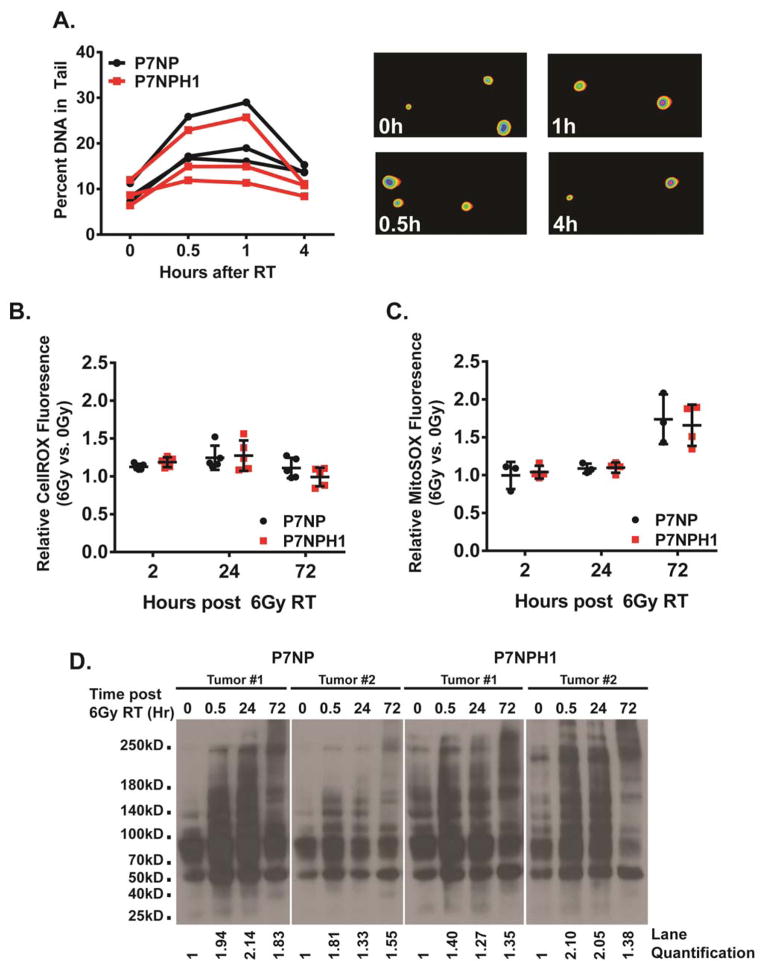
Figure 5. Deletion of HIF-1α did not affect DNA damage repair or cellular ROS clearance after irradiation.
Primary cells from P7NP and P7NPH1 tumors were isolated as described in Figure 3. All experiments were performed with in vitro cultures under 21% oxygen. A) DNA damage repair after 10Gy irradiation was measured with the Comet assay. Cells were irradiated and collected at indicated times, and embedded in agarose. DNA damage was measured by the percentage of visible tail after electrophoresis. Deletion of HIF-1α (P7NPH1) did not affect non-homologous end-joining repair as compared to sarcoma cells from P7NP mice by a two-tailed student t-test. Representative images of the DNA tail were shown in the right panels. B) Cells were irradiated with 6Gy. Accumulation of reactive oxygen species (ROS) was measured first by incubating nonirradiated and irradiated cells with CellRox DeepRed reagent at 2, 24, and 72 hours, then by performing flow cytometry. Relative ratios of ROS were calculated by normalizing the irradiated samples to nonirradiated controls at each time point. Deletion of HIF-1α (P7NPH1) did not affect ROS accumulation as compared to sarcoma cells from P7NP mice by a two-tailed student t-test. C) Cells were irradiated with 6Gy. Accumulation of mitochondrial superoxide was measured first by incubating nonirradiated and irradiated cells with MitoSOX reagent at 2, 24, and 72 hours, then by performing flow cytometry. Relative ratios of mitochondrial superoxide were calculated by normalizing the irradiated samples to nonirradiated controls at each time point. Deletion of HIF-1α (P7NPH1) did not affect mitochondrial superoxide accumulation as compared to sarcoma cells from P7NP mice by a two-tailed student t-test. D) Two P7NP and P7NPH1 primary sarcoma cell lines were irradiated with 6Gy. Damage to cellular structures by ROS was measured by protein carbonylation at indicated times after irradiation using OxyBlot per the manufacturer’s instructions. Deletion of HIF-1α (P7NPH1) did not affect the rate of accumulation or resolution of ROS-induced protein carbonylation. ImageJ quantification of cabonylated proteins for each time point was calculated as a ratio to the 0h time point, and listed below each lane.

