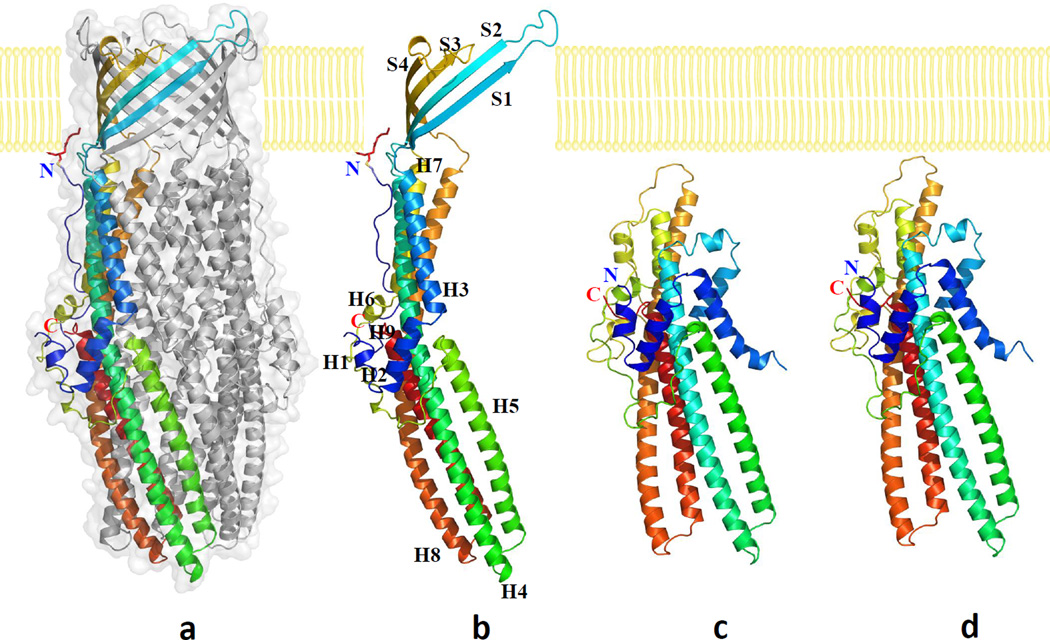
Fig. 1.
Structures of the CusC channel proteins. (a) Side view of the trimeric CusC channel. One of the protomers of CusC is in rainbow colors. The other two molecules of CusC are colored gray. (b) Ribbon diagram of the structure of a CusC protomer. The CusC protomer is acylated (red sticks) through the Cys1 residue to anchor onto the outer membrane. (c) Ribbon diagram of the structure of a protomer of the ΔC1 mutant. (d) Ribbon diagram of the structure of a protomer of the C1S mutant. The molecules are colored using a rainbow gradient from the N-terminus (blue) to the C-terminus (red).

