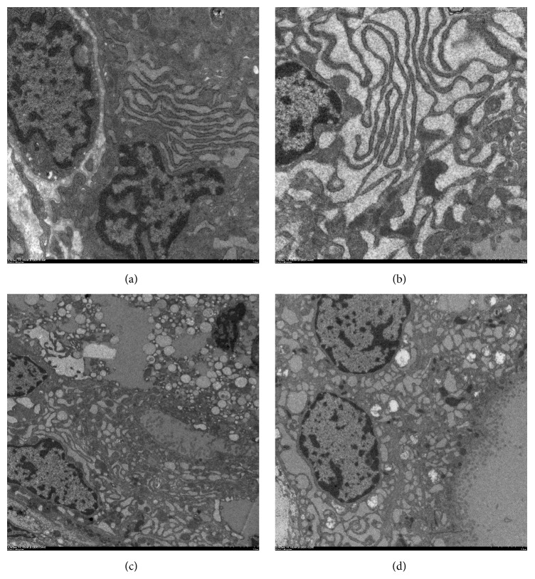
Figure 9.
TEM comparison of thyroid follicular cells architecture. (a) A TEM analysis from the control group showing that the follicular cells contain well-developed parallel cisternae of ER, mitochondria, and large moderate electron dense cytoplasmic colloid droplets. Apical lateral surfaces of follicular cells show tight junctions (magnification, ×5,000); (b) TEM analysis from the PCB group showed that the follicular cells contained many vacuoles and few microvilli. Follicular cells have euchromatic nuclei with peripheral rim of heterochromatin and markedly dilated cisternae of ER (magnification, ×5,000); (c) in PCB group, corrugated heterochromatic follicular cells nuclei and desquamated follicular cells within follicular lumen are noticed. The general loss of subcellular organization and cellular contents is also found (magnification, ×2,000); (d) TEM analysis from the PCB + APO group showing that the follicular cells cytoplasm contains moderately dilated cisternae of ER, Golgi saccules, and apical electron dense granules (magnification, ×2,000).

